
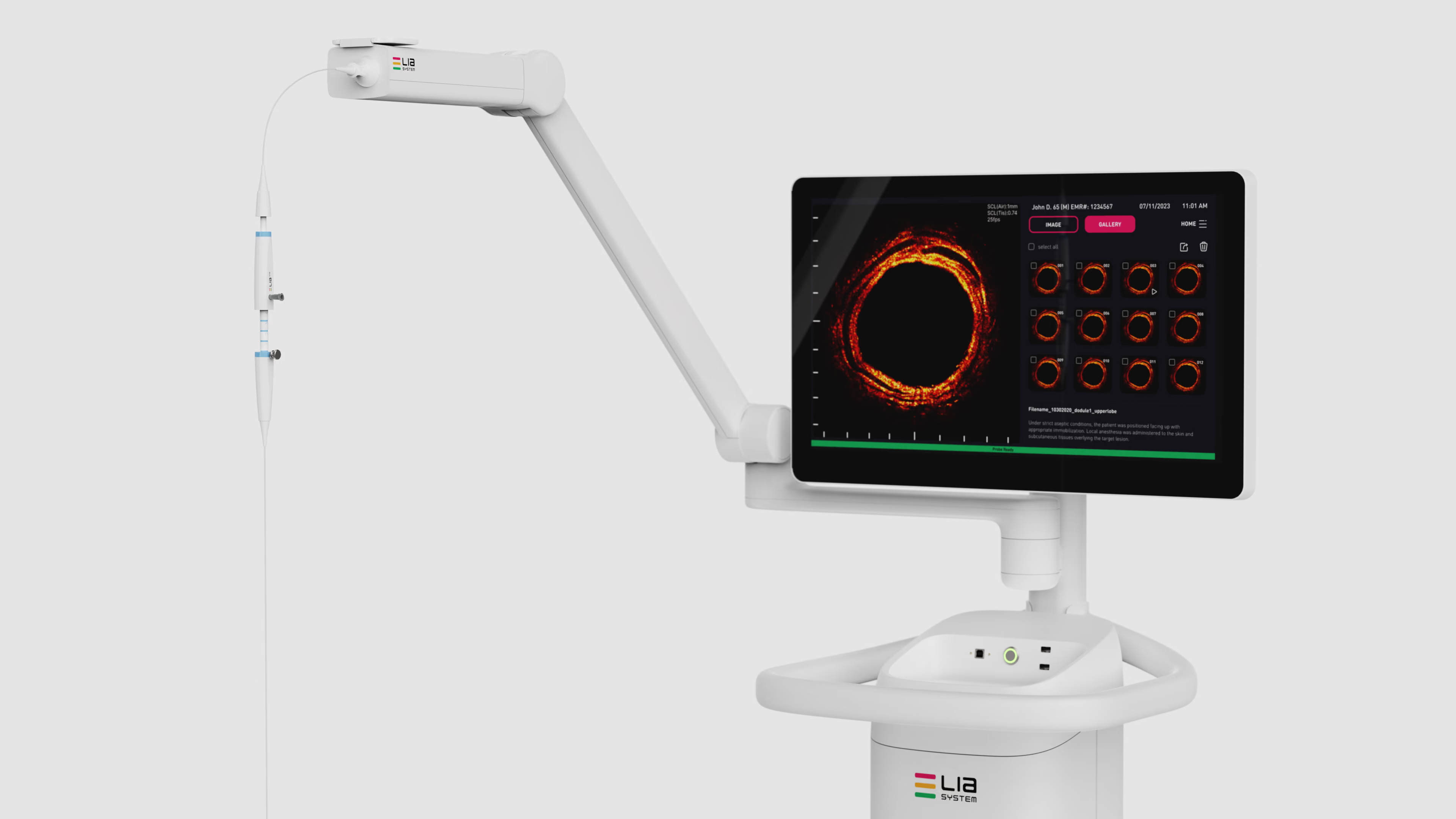
One Tool. One Continuous Workflow.
Image, guide, and sample through a single 21G needle, in real time.
Navigation ≠ Diagnosis.
Today's bronchoscopy platforms are exceptional at reaching peripheral lesions. The best systems achieve lesion access rates above 95%. Reaching the lesion is not the bottleneck. Confirming the tool is in it is.
LIA™ closes that gap by delivering real-time, in-tissue imaging at the exact moment of biopsy. Not proximity. Not navigation. Vision.
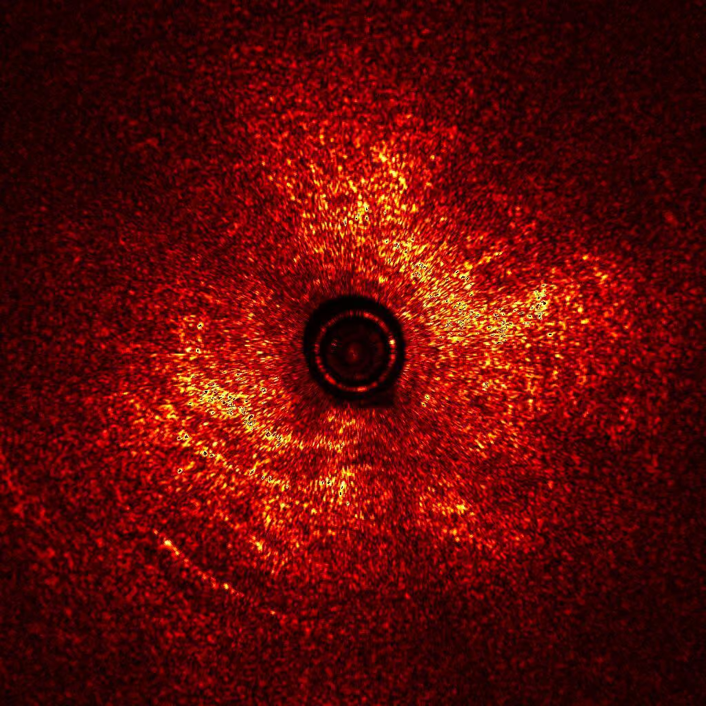
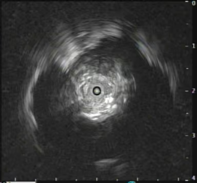
See through the noise.
Ultrasound cannot travel through air. It needs tissue contact or a coupling medium such as saline. LIA™ uses near-infrared light, which images across air and into tissue, with more than 10× the resolution of radial ultrasound. No breath-hold. No radiation. Signal remains stable through air, blood, and mucus. These are the real conditions of a live procedure.
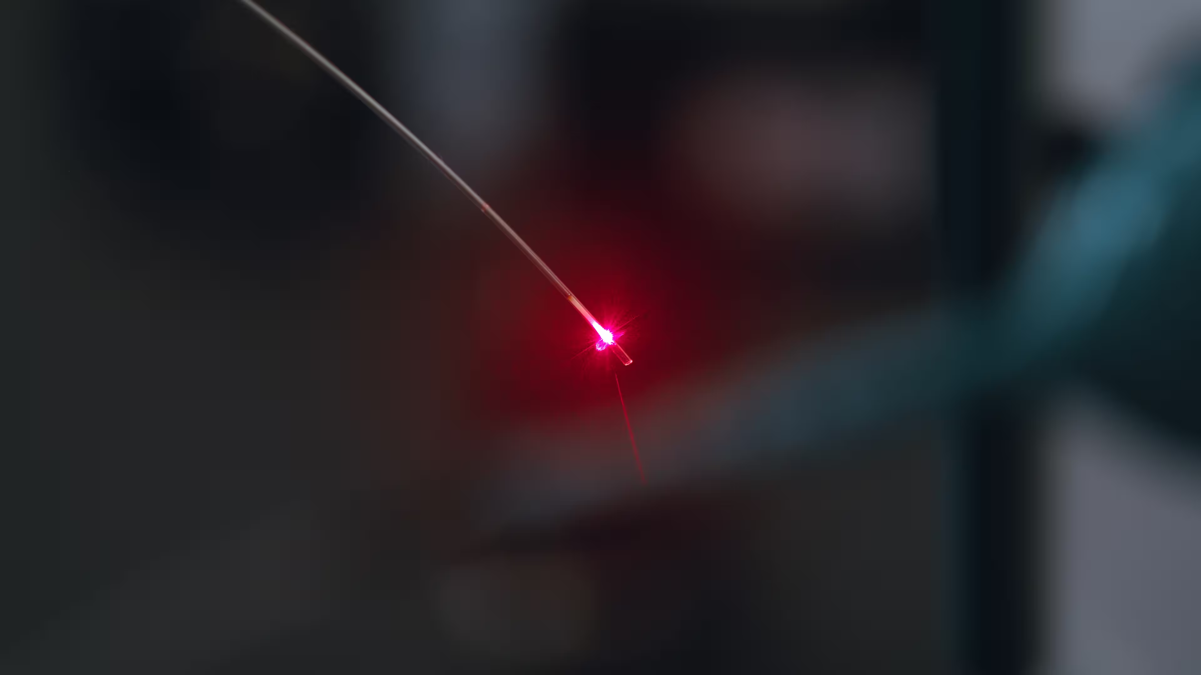
An Optical Chip Inside the Needle
Metasurface micro-optics, made with semiconductor processes, integrated into a 21G needle. Fits compatible bronchoscopes with a working channel of 1.7 mm or larger. 360° field of view.
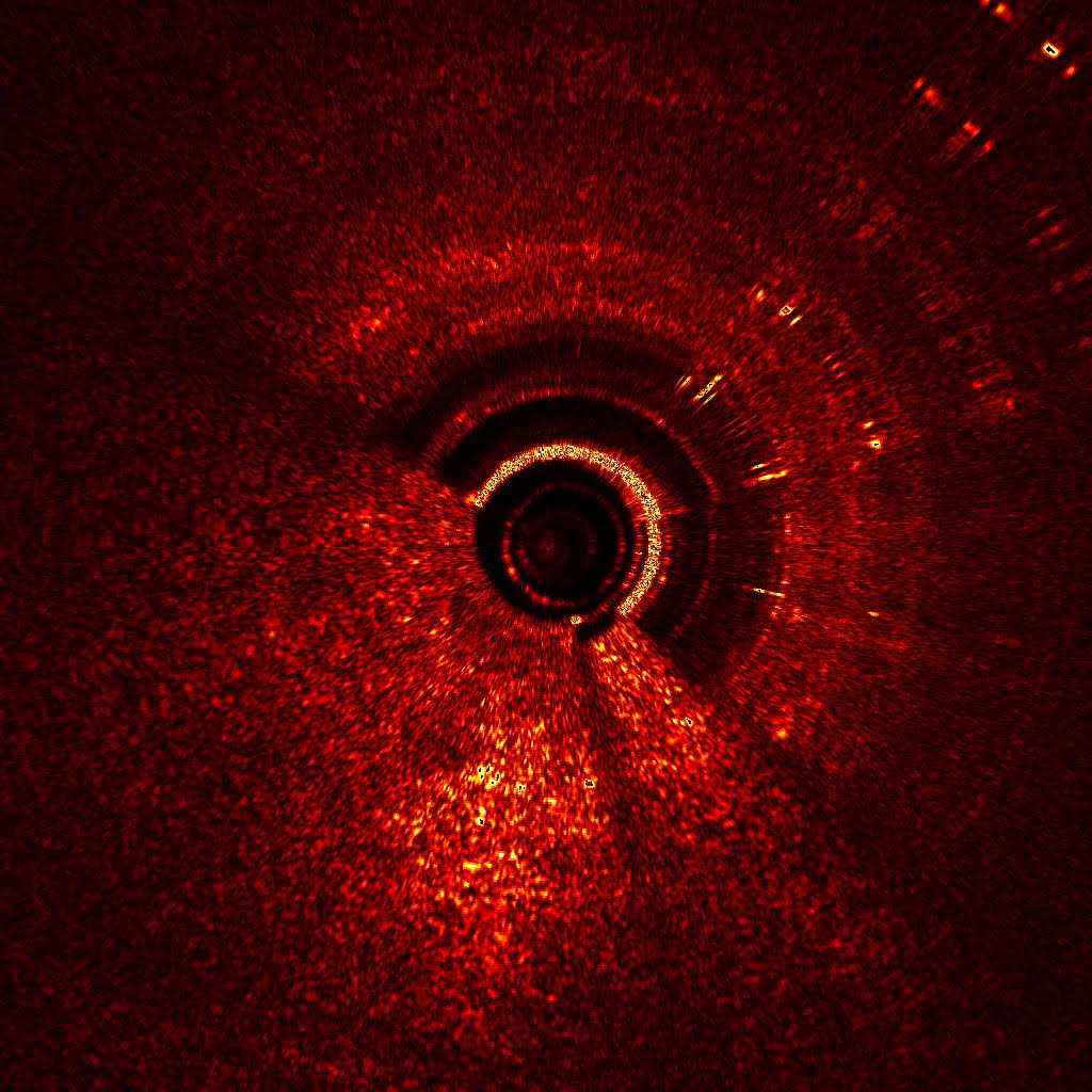
See the Tissue at the Needle Tip
LIA™ generates real-time, 360° cross-sectional images of tissue microstructure in the airway and lung parenchyma. The physician sees the tissue before taking the sample.
See it in action
How It Works in a Procedure
1
Navigate Use your existing bronchoscope. Robotic or traditional. LIA™ is platform-agnostic.
2
Deploy Advance the LIA™ catheter through the working channel to the target lesion.
3
Image Real-time near-infrared imaging shows tissue microstructure at the moment of biopsy. See the nodule. Know you're in it.
4
Sample One tool, one continuous workflow.
Ready to see it in your suite?